Understanding Degenerative Retinoschisis

This page provides comprehensive information on degenerative retinoschisis, detailing its nature, impact on vision, diagnostic methods, management strategies, and essential guidance for preserving retinal health. Associated Eye Physicians & Surgeons of NJ proudly offers eye care at four convenient locations: Belleville serving Essex County, Rahway serving Middlesex and Union Counties, Jersey City serving Hudson County, and Union serving Union County.
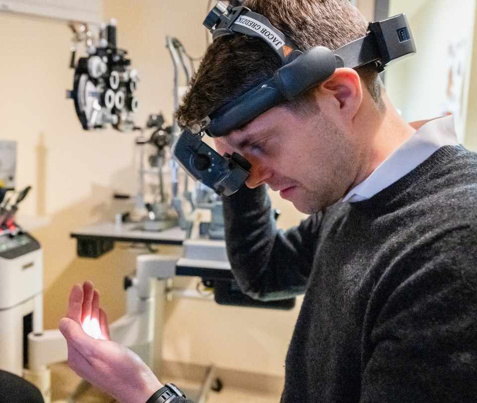
Dr. Bradford Carter Liva is a board-certified ophthalmologist specializing in retinal care, with a focus on injectable treatments for macular degeneration and diabetic retinopathy.
Overview of Degenerative Retinoschisis


Nature of the Split
In degenerative retinoschisis, the term “schisis” comes from the word for split or cleft. Essentially, a part of the retina may separate into two potential layers: one containing the light-sensing cells and the other containing the cells that relay signals to your brain via the optic nerve. The split can follow different patterns and is generally categorized by its appearance.
There are two primary subtypes described:
Impact on Vision
Our eyes work best when every layer of the retina is correctly aligned to capture light and create clear images. With degenerative retinoschisis, this organized structure is disrupted.
Many people with this form of retinoschisis may not notice any significant symptoms, primarily because the separation often occurs in areas of the retina away from your central vision. However, if the changes become more severe or if the condition affects the edges of your visual field, you might experience issues such as a loss of peripheral vision. In more pronounced cases, some patients describe seeing floaters, small spots or lines that drift in your field of vision, or flashes of light, which can be unsettling on their own.
It’s important to remember that even if you feel your vision hasn’t suffered dramatically, routine examinations can help catch subtle changes early. Our retina specialists stress the importance of these check-ups to ensure we address any potential progression of the condition.

Exploring the Causes
One of the puzzling aspects of degenerative retinoschisis is that its exact cause remains unclear. Unlike congenital forms of retinoschisis, where a genetic mutation is more evident, the acquired type doesn’t have a well-established origin in the medical community. Researchers continue to investigate why the retina splits, but what we do know is that the risk increases as we age.
This age-related association is why degenerative retinoschisis typically surfaces in people in their 50s, 60s, or 70s. As time goes on, the structural integrity of the vitreous—the gel-like substance in your eye—can change, which might contribute in small part to the onset of the condition. However, it is important to note that while age is a significant factor, not everyone in these age groups will experience degenerative retinoschisis. Factors such as the overall health of the retina and the natural aging process of the eye tissues also play roles.
While the medical community continues to piece together the exact mechanisms behind the retinal split, it is clear that current research emphasizes careful monitoring in at-risk populations to ensure any progression is caught early.


Diagnostic Methods
Because the symptoms of degenerative retinoschisis can be subtle, diagnosis is often made during a comprehensive eye exam conducted by our retina specialists. Several specialized tests are typically performed to look closely at the layers of the retina:
Differentiating Retinoschisis and Retinal Detachment
For many patients, understanding the difference between retinoschisis and retinal detachment is an important aspect of navigating their eye health. Though they may seem related, these two conditions affect the eye in distinct ways.
Retinal detachment occurs when the entire retina pulls away from the underlying supportive tissue. This can lead to a dramatic and immediate loss of vision, making it a sight-threatening emergency. In contrast, retinoschisis involves a separation within the retina itself, meaning that the layers split but the retinal tissue does not fully detach from its support. This key difference means that while retinal detachment often requires swift surgical intervention, degenerative retinoschisis might be observed over time with regular check-ups.
It is also possible, though less common, for a person to experience both conditions concurrently. When the bullous form of degenerative retinoschisis becomes more pronounced, the likelihood of forming retinal holes increases, and these holes can sometimes lead to a retinal detachment. However, early detection and monitoring can often prevent the transition from a benign split to a more severe detachment. If you’re ever uncertain about changes in your vision, don’t hesitate to discuss them with our retina specialists.

Management and Treatment
Strategies
While there is no cure for degenerative retinoschisis, many patients manage its progression through regular monitoring and practical interventions. The first step is always a thorough discussion with our retina specialists who can explain what any changes mean for your vision and how best to proceed.
In many cases, degenerative retinoschisis is considered benign, meaning that it does not significantly impair vision or require aggressive treatment. However, it is essential to remain vigilant, as a small percentage of cases can lead to complications like retinal holes or even retinal detachment. Here are some important considerations when managing this condition:
Frequently Asked Questions about Degenerative Retinoschisis
Confusion is natural when dealing with a retinal condition that isn’t broadly discussed. Here are some questions that patients commonly ask, along with straightforward answers:
Your Trusted Retina Specialists Serving Central and Northern NJ
Degenerative retinoschisis is a condition that can significantly impact your visual health if not monitored. Regular check-ups with our eye doctors are essential to track any changes and address potential complications. With proactive management, you can continue to lead a healthy, active life. At Associated Eye Physicians & Surgeons of New Jersey, we prioritize your retinal health and comfort. Experience personalized, expert eye care by scheduling an appointment with our eye doctors in Belleville, Jersey City, Rahway, or Union, NJ.
Request an Appointment
At Associated Eye Physicians of NJ, we’ve built our reputation on care that’s local, personal, and easy to get to. With offices in Belleville, Union, Jersey City, and Rahway, we’re proud to serve a wide range of communities across North and Central Jersey. Our Belleville team welcomes patients from all over Essex County, including Newark, Bloomfield, and Nutley. Rahway welcomes patients from across Middlesex and Union Counties, including nearby patients from Elizabeth, Avenel, Woodbridge, and Perth Amboy. Our Union office is a favorite for families from Union County, especially for those in Elizabeth and Cranford. And in Jersey City, we care for Hudson County residents, including our neighbors in Hoboken and Bayonne.
- Retinal Diseases
- Diabetic Retinopathy
- Juxtafoveal Telangiectasis
- Macular Degeneration
- Macular Holes
- Retinal Tears & Detachments
- Vascular Occlusions
- Sudden Vision Changes
- Uveitis
- Vitreous Hemorrhage
- Retina Treatments
- CRVO
- CSCR
- Choroidal Hemangioma
- Degenerative Retinoschisis
- Macular Pucker
- Plaquenil Toxicity
- Anti-VEGF
- Lowering AMD Risk
- Fundus Photography
- FFA
- Optical Coherence Tomography
- Eye Vitamins
- Driving With AMD
- Blood Glucose & Vision
- Early AMD Signs
- Living With AMD
- Reading With AMD
- Independent With AMD
- AMD & Blurry Vision
- Eye Floaters